

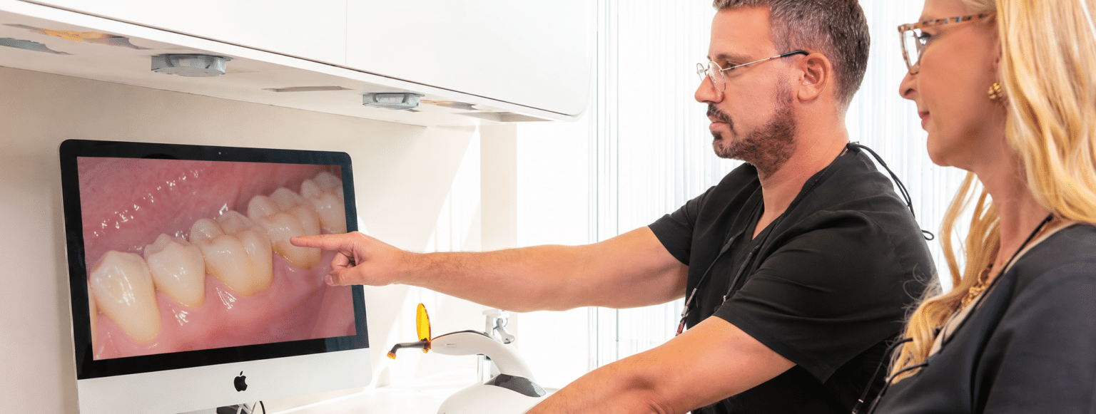
Intraoral & Fluorescence Camera
Patient communication, caries diagnostics and documentation in one.
“Insufficient MOD amalgam filling, 26 initial occlusal caries, approximal caries 25, secondary caries distal at the crown margin 46 and gingivitis on the lower front teeth…”
What does the dentist actually mean by that, and why do I have to have all of this treated now? My previous dentist said only a few months ago that everything was fine……….?

What your dentist sees — now you can see it too
Unfortunately, situations like this happen all too often and are only surprising in very extreme cases. The patient should not be left alone at this point; detailed information is necessary, and the findings should be visualised using the most modern diagnostic procedures currently available in dentistry so that the patient can understand them.

Detecting decay before it becomes visible
Whether for normal intraoral images or macroscopic magnified images, the VistaCam iX also supports the user in caries diagnostics. Here, the so-called Proof interchangeable head is used, which makes caries and plaque visible. Visualisation is made possible by fluorescence and, through numerical software analysis on the PC, also allows qualitative statements about the degree of the carious lesions. Just as with other diseases, the key to success here lies in prevention and early detection.
Our spaces are best experienced in person.
Arrange a no-obligation introductory visit — we’ll show you the practice, discuss your wishes and answer your questions.